Columbia University Irving Medical Center
Transforming human health by driving discovery, advancing care and educating leaders

About CUIMC
Our Schools
Advancing Care
News
- May 13, 2024
The annual Schaefer Scholar Awards are given to research scientists who have distinguished themselves in the science of human physiology and whose current work is of outstanding merit.
Topic
- May 9, 2024
Ever since Type A personality was linked to cardiovascular disease in the 1950s, it’s been known that anger raises the risk of heart attack and stroke. Now a Columbia study may explain how.
Topic
- May 1, 2024
Arthur G. Palmer and Oliver Hobert of the Department of Biochemistry & Molecular Biophysics were selected in recognition of their distinguished and continuing achievements in research.
Topic
- May 6, 2024
A new study reveals how bone marrow stem cell niche generation is regulated, which could lead to improvements in stem cell transplantation for the treatment of blood diseases.
Topic
- May 6, 2024
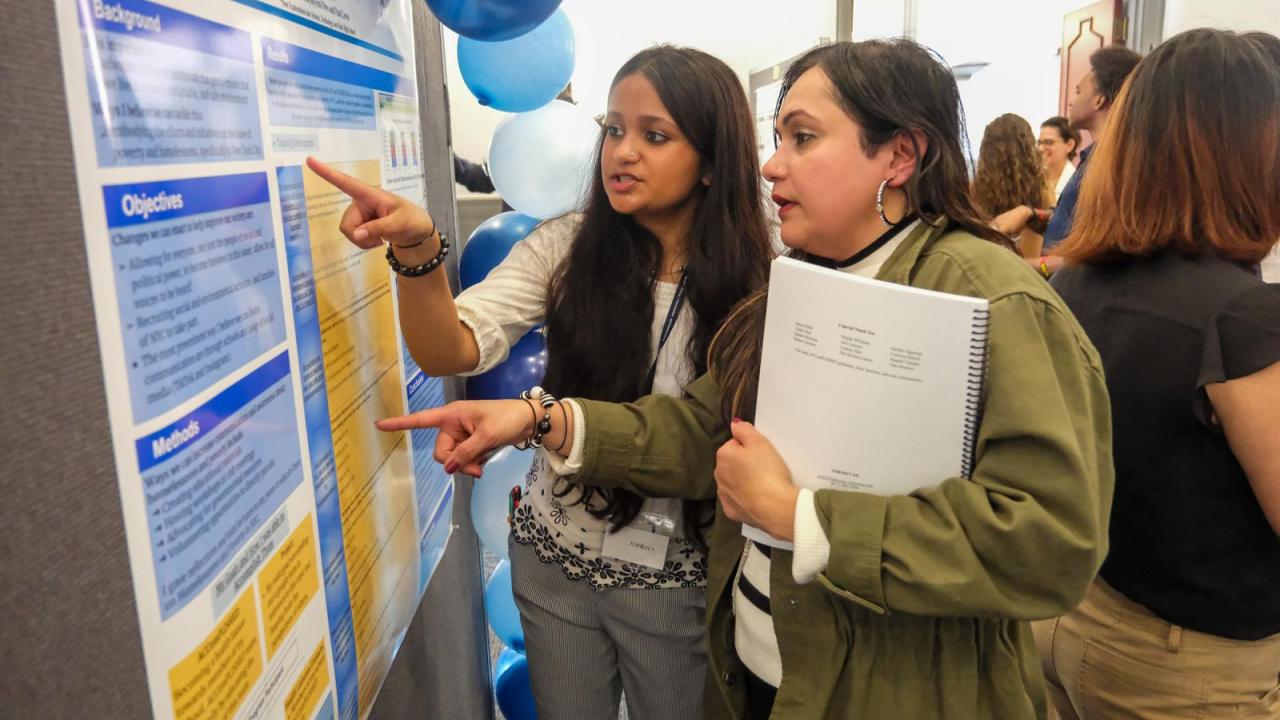
High school students who participated in a new community health education program created by a Columbia medical student celebrated their accomplishments at a graduation event.
Topic


Our Community

Learn more about our role in serving the Northern Manhattan community of Washington Heights, Inwood, and Harlem.
Diversity, Equity, and Inclusion
At CUIMC, we are committed to providing culturally inclusive medical education, research, and clinical care.
Events
- Thursday, May 16, 202411:30 AM to 12:30 PM
Venue
Hybrid Event - Thursday, May 16, 202411:30 AM to 12:30 PM
- Thursday, May 16, 202412:00 PM to 1:00 PM
- Thursday, May 16, 20244:00 PM to 5:00 PM
Venue
Online Event